Symptoms
The disease can appear approximately three weeks after planting. Symptoms consist of wilting, paler coloured leaves, shrunken stems above and below ground, followed by plants collapsing flat onto the ground. Infection of adult plants shows wilting progressing from the petioles and the youngest leaflets to the whole plant in two to three days. Leaves turn greyish green then a dull yellow colour, especially the lowest ones, followed by plant death. In both seedling and adult plant infections, inspection of roots split longitudinally shows a brown to black discolouration of internal vascular tissue (pith and xylem).
Chickpeas are infected via roots and the pathogen moves through the vascular system, produces enzymes that break down the host cells to form gels. These gels block the vascular system thus causing a yellowing and wilting of leaves and eventually plant death.
Organism
Fusarium oxysporum f.sp. ciceris
Host range
Chickpea (Cicer arietinum).
Method of spread
This pathogen can be spread with infected seed, soil, in plant debris, by water flow and by rain splash. It can survive in the soil for many years in the
absence of a host.
Conditions favouring disease
Conditions favouring disease are early planting. Disease development can be enhanced by failure to remove host debris from infected fields, shallow ploughing and crop rotations of less than three years.
Confused with?
Fusarium wilt of chickpea can be confused with phoma blight, damping off, rhizoctonia and phytophthora root rot, but may be distinguished from them by the discolouration of the internal root tissue.
Where?
India, Bangladesh, Myanmar, Nepal, North Africa, Spain and Mediterranean countries, America, Mexico and Brazil.
Thank you to Kurt Lindbeck (NSW DPI) for assistance with his contribution of content.
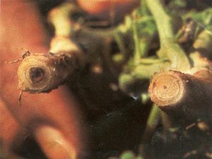
Image 2. Vascular discolouration (left stem). Source: M.P. Haware, Y.L. Nene & S.B. Mathur (ICRISAT)