You probably already have a good idea of what a Varroa mite looks like, even if you have never seen a hive with Varroa before. Based on photographs (Figure 1), you might expect to see a few tan/brown, hairy, oval-shaped attachments crawling on top of the bodies of worker bees. However, these images can be misleading; Varroa can be extremely difficult to identify by eye inside the hive, and might easily go unnoticed when infestation levels are low.
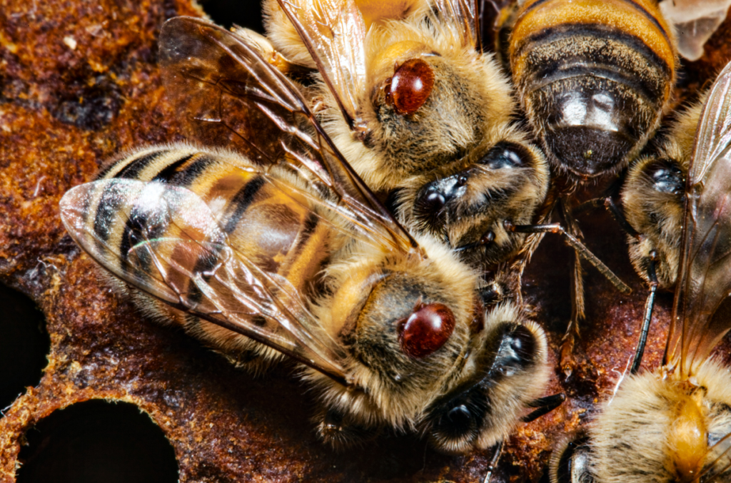
Figure 1. Mark Moffett / Minden Pictures
Figure 2. Mites are clearly visible to the human eye. Photo: Vincent Dietemann.
Mites are just over 1 millimetre in size – about the same size as a poppy seed. Considering a honey bee body is about 5mm wide (minus the wings), mites take up a lot of space on a bee thorax (Figure 1). Although poppy seeds (and Varroa mites) are visible to the naked eye (Figure 2), it is rare to see a mite perched neatly on the thorax of a bee. More than 80% of the time, mites are wedged deeply within the hard outer layers of the underside of the bee’s abdomen (the ‘tergites’; Figure 3), where they can be found feeding on the ‘fat bodies’ of bees, the fatty organ that is the equivalent of the insect liver. Mite and bee exoskeletons have similar pigmentation, so the mites are nicely camouflaged by their matching colours.
Not all mites are red/brown (Figure 4, 5). Mature females stand out better, but young mites vary in colour from white to tan as they mature (Figure 6). Males are white or white/yellow and are circular in shape (Figure 6).
Mites spend most of their time in the brood, so you are more likely to find them there (Figure 7). There are various ways to conduct surveillance, including alcohol or soapy water washes, sugar shake, brood uncapping and sticky boards.
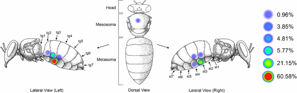
Figure 3. Varroa destructor shows consistent preference for the underside of the metasoma of adult host bees, an area predominated by fat body tissue just beneath the cuticle. (Left) Diagram showing frequency of Varroa found in each location on 104 parasitized worker bees in five trials (st, sternite; tg, tergite). Ramsey et al (2019)
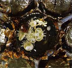
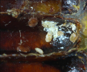
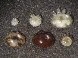
Figure 6. Upper left row from left to right: protonymph, deutonymph, deutochrysalis. Lower row from left to right: freshly moulted young female, mother mite, adult male. Rosenkranz et al 2010
Above left: Figure 4. A mite family with mother mite (reddish brown) and different stages of offspring at the bottom of a cell from which the honey bee pupa was removed. Photo: Denis Anderson. | Above right: Figure 5. In this section of a cell (the bottom is on the right side), the pearly white faeces deposit is visible on the upper and back walls. Mature and immature varroa mites are also visible. Photo: Swiss Bee Research Institute
The life cycle of varroa
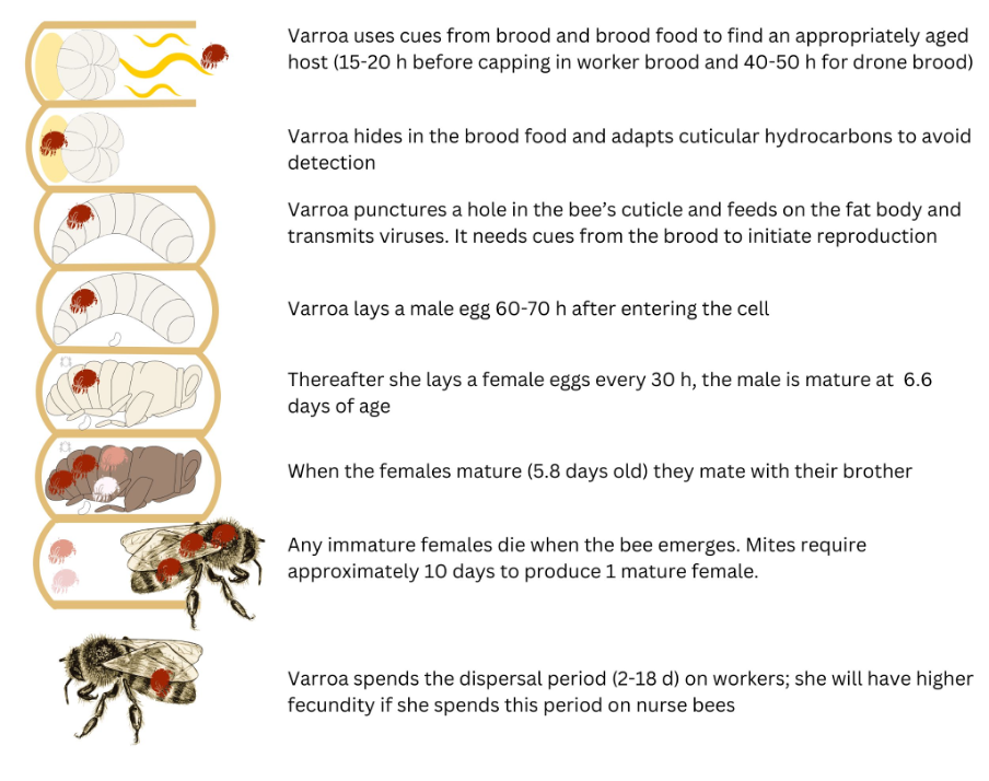
Figure 7. The life cycle of Varroa – Resilient beekeeping in the face of Varroa
Video: Amazing Time-lapse: Bees hatch before your eyes | National Geographic
Acknowledgements:
- Resilient beekeeping in the face of Varroa. AgriFutures Australia. Holmes, Gerdts, Grassl, Mikheyev, Roberts, Remnant and Chapman.
- Slowing the spread. AgriFutures Australia and AHBIC. Holmes, Gerdts, Grassl, Mikheyev, Roberts, Remnant and Chapman.
- Bee Aware
- Dietemann et al (2013) Standard methods for varroa research. Journal of Apicultural Research 52: 1-54
- NSW DPI Primefact – Varroa mites (2022)
- Ramsey et al (2019) Varroa destructor feeds primarily on honey bee fat body tissue and not hemolymph. PNAS 116: 1792-1801
- Rosenkranz et al (2010) Biology and control of Varroa destructor. Journal of Invertebrate Pathology 103: S96-S119
- This article was peer-reviewed by Michael Holmes and Mark Page.